
Digital X-Ray
Digital X-ray technology provides high-resolution images with reduced radiation exposure compared to traditional X-rays. This advanced imaging technique offers quick and accurate diagnostics, allowing for faster detection and treatment of conditions. Ideal for various medical assessments, digital X-rays enhance patient safety and streamline the diagnostic process.

Ultrasound Antenatal Scan
An ultrasound antenatal scan is essential for tracking fetal development during pregnancy. Using sound waves, this non-invasive test produces detailed images of the fetus and uterus, enabling healthcare providers to assess growth, detect anomalies, and ensure both maternal and fetal health. Regular scans support early diagnosis and informed care.
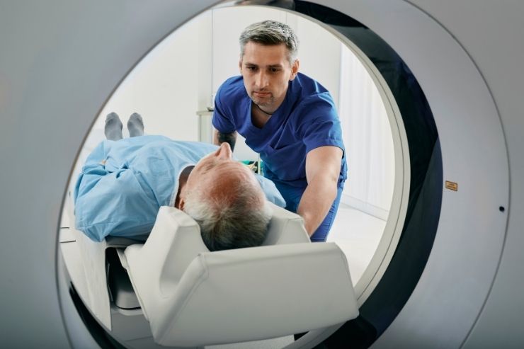
CT Scan
A CT scan, or computed tomography scan, uses X-rays and computer technology to produce detailed cross-sectional images of the body. This advanced imaging technique provides comprehensive views of internal structures, aiding in the diagnosis of various conditions such as tumors, internal injuries, and diseases. CT scans offer quick, precise results, enhancing diagnostic accuracy and guiding effective treatment plans.
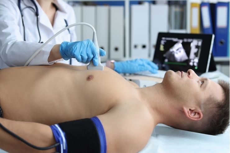
Echocardiography / Stress Echocardiography
Echocardiography is a diagnostic test that uses sound waves to create detailed images of the heart's structure and function. This non-invasive procedure helps assess heart chambers, valves, and blood flow, enabling the detection of abnormalities such as heart disease, valve disorders, and congenital defects.

Anomaly Scan
An anomaly scan, typically conducted between 18-22 weeks of pregnancy, provides detailed imaging of the fetus to identify potential structural abnormalities. This crucial ultrasound examines the baby’s organs, limbs, and overall development, enabling early detection of congenital issues and guiding appropriate prenatal care and intervention.
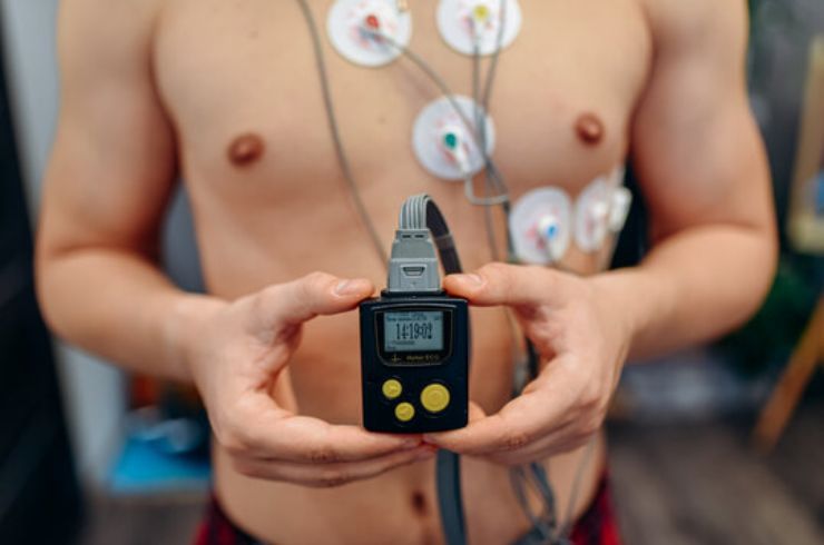
TMT, Holter & PFT
A Treadmill Test (TMT) evaluates the heart’s performance during exercise to detect coronary artery disease and irregular rhythms. Holter monitoring records heart activity over 24-48 hours, identifying arrhythmias and palpitations. Pulmonary Function Tests (PFT) assess lung capacity and airflow, aiding in the diagnosis and management of respiratory conditions.
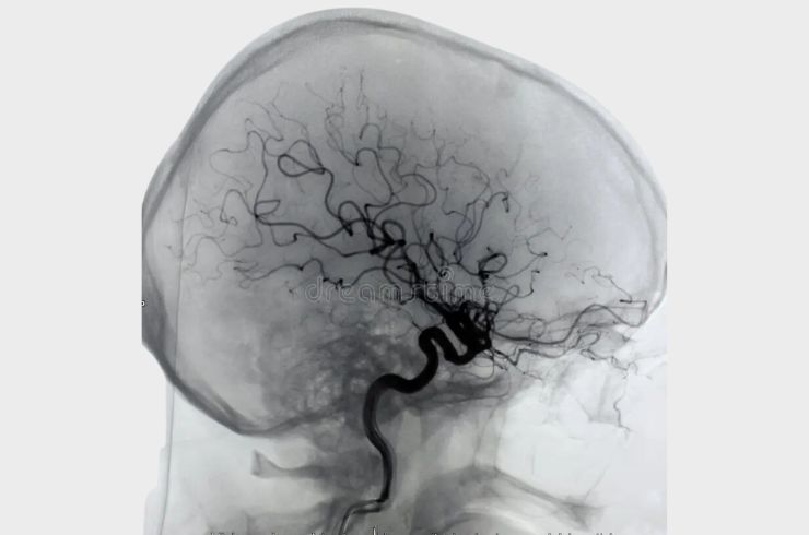
Fluoroscopy, Digital Subtraction Angiography
Fluoroscopy provides real-time X-ray imaging to visualize moving structures, aiding in diagnostics and procedures like catheter placements. Digital Subtraction Angiography (DSA) enhances vascular imaging by removing overlapping structures, offering detailed visuals of blood vessels. These advanced techniques are essential for diagnosing conditions and guiding minimally invasive treatments with precision.
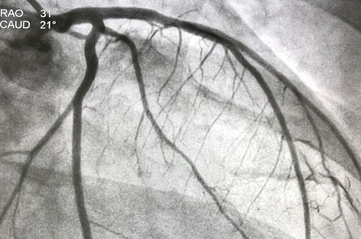
Coronary Angiography
Coronary Angiography is a specialized imaging procedure that uses contrast dye and X-rays to visualize the coronary arteries. It identifies blockages or narrowing that may impede blood flow to the heart. This essential diagnostic tool aids in detecting coronary artery disease and guides effective treatment strategies, including angioplasty or bypass surgery.


